
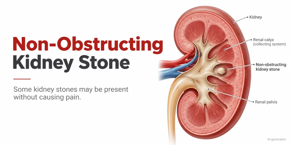
A kidney stone can sometimes be discovered during an ultrasound even when a person has no pain or urinary symptoms. This is called an incidental kidney stone, meaning the stone was identified during an imaging examination performed for another medical reason.
An incidental finding does not automatically mean that treatment is necessary. However, the stone’s size, position, potential to obstruct urine flow and the person’s medical history may influence whether additional evaluation or monitoring is appropriate.
An incidental kidney stone may be detected during an imaging examination performed for another reason. Learn more about kidney and ureteric stones and their possible symptoms and management options.An incidental kidney stone is a solid deposit of minerals and salts found in the kidney during an examination that was not specifically intended to investigate kidney stone symptoms.
For example, a person undergoing abdominal ultrasonography for a digestive concern or a routine evaluation may receive a report mentioning a small renal calculus.
The term asymptomatic kidney stone means that the stone is not currently producing noticeable symptoms.
Some kidney stones remain within the kidney without causing obstruction. Others may grow, move into the ureter or cause symptoms later. The future behaviour of an individual stone cannot always be predicted.
Ultrasound uses sound waves to create images of internal organs, including the kidneys.
During the examination, certain stones may appear as bright structures that reflect sound waves. Some also produce an acoustic shadow behind them, helping the examiner identify a possible calculus.
Ultrasound may provide information about:
The approximate size and location of a kidney stone.
Whether one or both kidneys contain visible stones.
Possible swelling of the kidney due to impaired urine drainage, called hydronephrosis.
Other structural findings that may require evaluation.
Ultrasound does not involve ionising radiation, making it useful in many clinical situations.
However, it has limitations. Very small stones or stones in certain locations may be missed. Ultrasound measurements can also differ from measurements obtained using CT imaging.
Kidney stone pain commonly occurs when a stone moves into the ureter and interferes with normal urine drainage.
A stone remaining within the kidney may not produce significant discomfort, particularly when it is not obstructing urine flow.
Several factors can influence symptoms:
Stone location: Stones within the kidney may behave differently from stones in the ureter.
Urinary obstruction: Blockage can increase pressure within the urinary collecting system.
Stone movement: Movement into the ureter may trigger sudden pain.
Associated infection: Infection can produce additional symptoms and may require urgent assessment.
The absence of pain does not always indicate that a stone is harmless. Clinical evaluation is important when imaging identifies a new or unexplained finding.
Not every incidental kidney stone represents an immediate health concern.
Some small, non-obstructing stones remain stable for extended periods. Others may increase in size, cause pain, contribute to infection or interfere with urine drainage.
| Factor | Why It Matters |
|---|---|
| Stone Size | May influence the likelihood of growth, passage and possible intervention. |
| Stone Location | Helps assess drainage and potential future movement. |
| Hydronephrosis | May indicate impaired urine flow requiring further evaluation. |
| Infection | Infection associated with obstruction can become a medical emergency. |
| Previous Stone History | Recurrent stones may justify additional prevention assessment. |
| Kidney Function | Reduced function or a solitary kidney may require closer attention. |
Important: A healthcare professional considers these findings together rather than relying on stone size alone.
Additional investigations are not necessary for every patient. The decision depends on the ultrasound report, symptoms, medical history and clinical examination.
A urinalysis may help identify blood, signs of infection or other urinary abnormalities.
If infection is suspected, a urine culture may also be requested.
Blood tests may assess kidney function, including serum creatinine. Selected metabolic tests, such as calcium measurements, may be appropriate depending on the clinical situation.
A non-contrast CT scan can provide more detailed information about stone size, number and location.
However, CT exposes patients to ionising radiation. It is not automatically required after every incidental ultrasound finding.
People with recurrent stones or particular risk factors may benefit from further assessment, sometimes including a 24-hour urine collection.
The aim is to investigate factors that may contribute to stone formation and recurrence.
No. An asymptomatic, non-obstructing kidney stone does not always require immediate removal.
For selected patients, active surveillance may be appropriate. This involves monitoring the stone and reassessing the situation if symptoms, imaging findings or other clinical factors change.
The 2026 American Urological Association guidance recognises both active surveillance and preventive surgical intervention as options for selected people with asymptomatic, non-obstructing kidney stones. Decisions should consider the available evidence, individual circumstances and patient preferences.
Treatment may be considered when a stone grows, causes obstruction or symptoms, is associated with certain infections, or presents other clinically important risks.
Procedures such as shock wave lithotripsy or ureteroscopy also have possible complications. Their benefits and limitations need to be discussed before a decision is made.
When observation is selected, a clinician may recommend periodic imaging to assess whether the stone remains stable.
The monitoring plan may consider:
Changes in stone size or number.
Development of pain or urinary symptoms.
Signs of infection or obstruction.
Kidney function and relevant metabolic risk factors.
There is no single follow-up schedule suitable for everyone. Ultrasound is often useful for surveillance, while other imaging may be considered when additional information is required.
Dietary recommendations may vary according to stone composition and individual health conditions. Learn about dietary considerations for kidney stones and approaches to recurrence prevention.General preventive measures may help reduce the risk of future stone formation, although recommendations depend partly on stone type and individual health conditions.
Adequate fluid intake can help reduce urine concentration. A balanced diet with appropriate calcium intake, moderate sodium consumption and attention to excessive animal protein intake may also be relevant.
People with kidney disease, heart failure or prescribed fluid restrictions should follow their clinician’s individual recommendations.
Dietary changes cannot be assumed to dissolve an existing kidney stone. Certain stone types, such as uric acid stones, may respond to specific medical treatment under professional supervision.
A newly discovered kidney stone should be discussed with a qualified healthcare professional to determine whether further evaluation is required.
Seek urgent medical attention when symptoms include:
Fever or chills with suspected kidney stone symptoms.
Severe or persistent pain in the side or back.
Repeated vomiting or inability to maintain fluid intake.
Difficulty passing urine or markedly reduced urine output.
Symptoms of a stone in someone with a solitary kidney or known kidney impairment.
A suspected obstructing stone with infection requires urgent assessment because delayed treatment can lead to serious complications.
Yes. Some kidney stones remain inside the kidney without causing noticeable pain or urinary symptoms. These are often described as asymptomatic kidney stones.
Ultrasound can identify some small stones, but its accuracy varies. Very small stones may be missed, and measurements may not always match CT findings.
No. Some asymptomatic, non-obstructing stones can be monitored. The decision depends on stone characteristics, clinical risk factors and individual preferences.
Yes. A kidney stone may increase in size while remaining asymptomatic. Follow-up imaging may help identify changes over time.
Not always. CT may be considered when the diagnosis is uncertain, symptoms develop, obstruction is suspected or detailed imaging is needed for treatment planning.
Some stones may eventually move into the ureter and pass through the urinary tract. However, spontaneous passage cannot be predicted reliably for an individual kidney stone.
An incidental kidney stone is a stone discovered during imaging performed for another reason, often before any symptoms develop.
Although many asymptomatic, non-obstructing kidney stones can be monitored, some may grow or cause complications. Understanding the ultrasound findings, assessing individual risk factors and selecting an appropriate follow-up plan can support informed decisions.
Medical Reviewer: Dr. Prakash Sankapal, Urologist — pending clinical review and approval.
Medical Disclaimer: This article is intended for general educational purposes only. It does not replace individual medical advice, diagnosis or treatment. Investigations and management should be determined by a qualified healthcare professional based on the patient’s medical condition.
National Institute of Diabetes and Digestive and Kidney Diseases (NIDDK). Diagnosis of Kidney Stones. https://www.niddk.nih.gov/health-information/urologic-diseases/kidney-stones/diagnosis
European Association of Urology (EAU). Guidelines on Urolithiasis, 2026. https://uroweb.org/guidelines/urolithiasis/chapter/guidelines
American Urological Association (AUA). Surgical Management of Kidney and Ureteral Stones: AUA Guideline, 2026. https://pubrica.com/wp-content/uploads/2026/02/The-Journal-of-Urology_AUA-Guideline-Article.pdf
Medical disclaimer: This article provides general educational information and does not replace personalised medical advice, diagnosis or treatment. Kidney mass assessment and management require review of individual clinical findings.
Plot no 70, Moreshwar Heritage, Sector 5, Ulwe, Navi Mumbai, Maharashtra 410206
For Booking: +91 8591009591
Chat With Us
49, Sion – Panvel Hwy, Sector 7, Vashi, Navi Mumbai, Maharashtra 400703
For Booking: +91 8007542142
Chat With Us